CTs and MRIs use different technologies to show different things — neither is necessarily ’better’ than the other

Image content: This image is available to view online.
View image online (https://assets.clevelandclinic.org/transform/dc77da66-ca46-4223-a36f-e3d868887f2d/MRI-versus-CT-Scan-MLC-967x544-1_jpg)
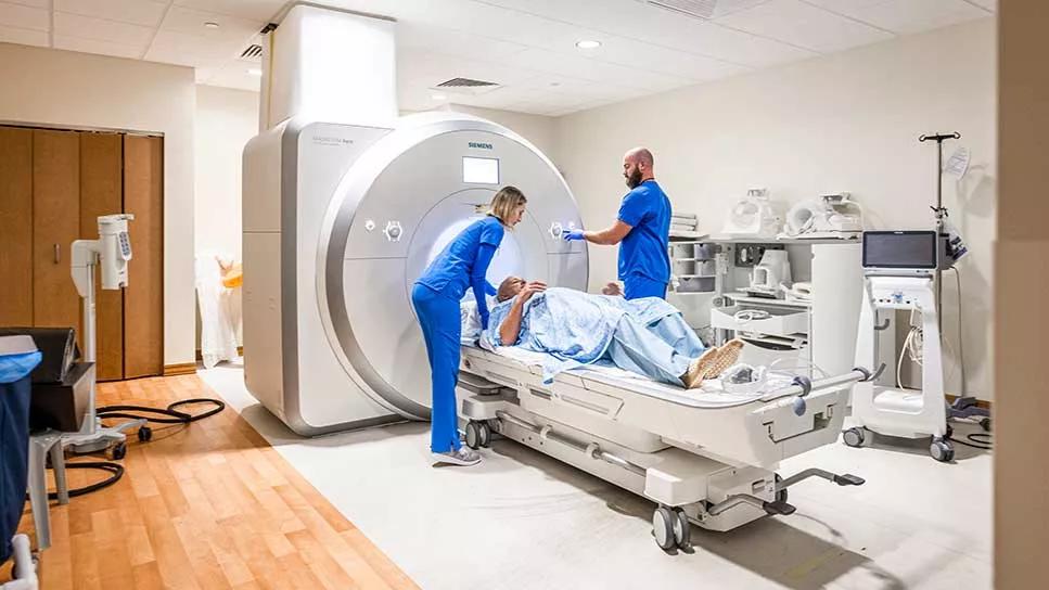
patient lying on imaging table about to enter machine
Some injuries or conditions you can see with the naked eye. Others require a more in-depth look.
Advertisement
Cleveland Clinic is a non-profit academic medical center. Advertising on our site helps support our mission. We do not endorse non-Cleveland Clinic products or services. Policy
And if your provider suspects a condition like internal bleeding, tumors or muscle damage, they may order a CT scan or MRI.
The choice to use a CT scan vs. an MRI is a call your provider makes, primarily based on what they suspect they’ll find.
“MRIs and CTs are two ways of looking inside your body, beyond the skin, and seeing what’s going on,” explains radiologist Albert Parlade, MD. “MRIs and CTs are able to see different things inside your body to help your provider make a diagnosis. They each work differently in different parts of the body.”
How do CTs and MRIs work, and which is best for what? Let’s take an in-depth look.
CT scans and MRIs use different technologies to see what’s happening inside your body. They’re imaging technologies, not treatments. That means they help your provider make diagnoses, plan your treatment and monitor changes in your body.
Both CT and MRI help your provider see structures inside your body. But which test is better for you depends on certain factors, like what exactly the provider is searching for, as well as your health status.
For a quick look, here are some differences between CT and MRI:
| CT | MRI | |
|---|---|---|
| Technology | X-ray. | Magnets and radio waves. |
| Examples of uses | Bones, stones, blood, organs, lungs, cancer stages and abdominal emergencies. | Joints, nerves, brain and masses. |
| Risks | Potential for radiation exposure. | Not for people with some metal implants or who can’t lie still for the procedure. |
| Procedure time | Less than a minute. | 20 to 50 minutes. People must lie very still throughout. |
| CT | ||
| Technology | ||
| MRI | ||
| X-ray. | ||
| Magnets and radio waves. | ||
| Examples of uses | ||
| MRI | ||
| Bones, stones, blood, organs, lungs, cancer stages and abdominal emergencies. | ||
| Joints, nerves, brain and masses. | ||
| Risks | ||
| MRI | ||
| Potential for radiation exposure. | ||
| Not for people with some metal implants or who can’t lie still for the procedure. | ||
| Procedure time | ||
| MRI | ||
| Less than a minute. | ||
| 20 to 50 minutes. People must lie very still throughout. |
Now, we’ll take a look at both to better understand what they’re best at and who they’re best for.
Advertisement
CT stands for “computed tomography scan.” Some people also call it a CAT scan.
You can think of a CT scan as a 3D (three-dimensional) X-ray machine.
“A CT scanner uses an X-ray that shoots through the patient to a detector as it spins around the patient,” Dr. Parlade explains. “It takes hundreds of pictures that a computer then puts together to create a 3D picture of the patient. You can slice and dice those images in different ways to get views inside the body.”
A traditional X-ray can give your provider one look at the area that was images. It’s a static photo.
But you can look at CT images to get a bird’s eye view of the area that was imaged. Or spin around to look from front to back or side to side. You can look at the outermost layer of the area. Or zoom deep inside of the part of the body that was imaged.
Getting a CT scan should be a quick and painless process. You lie on a table that moves slowly through a donut-shaped scanner. Depending on what your provider is looking for, you may also need to have an IV injection of a contrast dye. Each scan takes less than a minute.
Because CT scanners use X-rays, they can show the same things as an X-ray but with much more precision. An X-ray is a flat look at the area being imaged, whereas a CT gives a more complete, in-depth picture.
CT scans are used to look at things like:
“CTs are good at looking ‘bones and stones,’ like kidney stones and gallstones,” Dr. Parlade says. “Also, surgeons commonly use CTs to plan surgeries. For example, an orthopaedic surgeon may want a 3D image of a bone before repairing a complex fracture.”
CT scans also can be used to see things that MRIs don’t see well, like your lungs, blood and bowels. More on that in a bit.
The biggest concern some people have with CT scans (and X-rays for that matter) is the potential for radiation exposure.
“X-rays use a small amount of ionizing radiation, which is thought to possibly carry a risk for people. So, we’re cognizant of that when we consider whether someone should get a CT,” Dr. Parlade notes.
Some experts suggest that the ionizing radiation given off by CT scans can slightly increase cancer risk in some people. But the exact risk is a matter of controversy. The U.S. Food and Drug Administration says the risk of cancer from CT radiation is “of statistical uncertainty” based on current scientific knowledge.
But because there may be some risk from CT radiation, pregnant people are usually not good candidates for CT scans unless necessary.
Advertisement
And there are times when a provider may decide to use an MRI over CT to lower any risk of radiation exposure. That’s especially the case for people whose health conditions will require multiple rounds of imaging over long periods of time.
“An example is people living with Crohn’s disease,” Dr. Parlade illustrates. “They’re usually young people who are going to need multiple scans over their lifetimes. In those cases, we typically choose MRI over CT scan because it’s believed that accumulated radiation over their life can add up to increased risk.”
MRI stands for “magnetic resonance imaging.” In short, MRIs use magnets and radio waves to create images inside your body.
The exact way it works includes a long lesson in physics. But in short, it goes a little something like this:
Our bodies contain a lot of water, or H20. The H in H20 stands for hydrogen. Hydrogen contains protons — positively charged particles.
Typically, those protons spin around in all different directions. But when they encounter a magnet, like in an MRI machine, those protons get pulled toward that magnet and start to line up.
Then, come the radio waves. They give the protons a push and they start to spin. When the radio waves turn off, the protons go back to lining up to the magnets. As they get back in line, they send off signals.
Advertisement
The MRI machine translates those signals to create images of the inner workings of your body.
An MRI is a tube-shaped machine. A typical MRI scan takes about 30 to 50 minutes and you have to lie very still during the procedure. The machines can be loud, and some people may benefit from wearing earplugs or using headphones to listen to music during their scan. Depending on what your provider is looking for, they may use IV contrast dye.
MRIs are really good at differentiating tissue. For example, a provider may use a full-body CT to hunt for tumors. Then, order an MRI to better understand any masses found in the CT.
Your provider can also use an MRI to look for joint damage and nerve damage.
“You can see some nerves with MRI. You can see if there’s injury or inflammation in a nerve in certain parts of the body,” Dr. Parlade states. “We don't directly see nerves on CT scan. On a CT, we can see the bones around nerves or tissues around nerves and see if they’re doing anything to the area where we expect the nerve to be. But for directly seeing the nerves, MRI is a better test.”
MRIs aren’t as good at seeing some other things, like bones, blood, lung and bowels. Remember that MRIs in part depend on using magnets to affect the hydrogen in water that’s in your body. So, dense things, like kidney stones and bones, don’t show up as well. Neither do things full of air, like your lungs.
Advertisement
“Things like your appendix and bowels may not show up well on an MRI,” Dr. Parlade clarifies. “The MRI takes a few minutes to acquire. And those organs can move a lot, so they may not show up clearly.”
While an MRI may be a better technique to see certain structures in your body, not everyone is a good candidate.
If you have certain types of metal in your body, you can’t get an MRI. That’s because an MRI is essentially a magnet, so it can interfere with certain metal implants. That includes things like some pacemakers, defibrillators or shunts.
“If you have a metal implant, we have to check the label of those implants to make sure they're safe for MRI,” Dr. Parlade explains.
The metal in things like joint replacements is typically MRI-safe. But be sure your provider is aware of any metal in your body before you do an MRI scan.
Additionally, MRIs require you to stay very still for a period of time, which some people can’t tolerate. For others, the enclosed nature of an MRI machine can trigger anxiety or claustrophobia, which can make it very difficult to get imaged.
Neither a CT nor an MRI is always better — it’s a matter of what you’re looking for and how well you can tolerate each, Dr. Parlade emphasizes. “A lot of times, people think one is better than the other. But it really depends on what your doctor's question is.”
Bottom line: Whether your provider orders a CT or an MRI, the goal is to see what’s happening inside your body to get you the best treatment.

Sign up for our Health Essentials emails for expert guidance on nutrition, fitness, sleep, skin care and more.
Learn more about our editorial process.
Advertisement
One medical test, two different parts
Understand how these imaging tests differ
Bleeding is a risk and warrants taking care, but the reward of this lifesaving medication is great
Severe and debilitating headaches can affect the quality of your child’s life
With repeat injections over time, you may be able to slow the development of new wrinkles
Type 2 diabetes isn’t inevitable with these dietary changes
Applying a hot or cold compress can help with pain
Pump up your iron intake with foods like tuna, tofu and turkey